Critical Care in Patient with Neuromuscular Cervicothoracic Kyphosis
Article information
Abstract
A 10-year-old boy had a neuromuscular cervicothoracic kyphosis and kyphotic deformity got worse as he grew. He underwent posterior spinal fusion from T6 to pelvis two years ago. However, kyphosis progressed gradually, and difficulty occurred in breathing with a ventilator. We perform deformity correction with vertebral column resection at T5 and posterior fixation from T2 to T9 and posterior onlay fusion. Surgical correction is offered to stop the kyphosis progression, and finally to maintain the airway. After surgery, the patient transferred to an intensive care unit for respiratory care. The patient's breathing was much better than before surgery, and the patient was transferred to the general ward. We report the importance of postoperative care in spinal deformity patient with respiratory distress.
INTRODUCTION
Spinal deformity surgery in children with neuromuscular spinal deformity (NSD) is associated with a high morbidity rate1,4,8-10). Respiratory complications following spinal deformity surgery in patients with NSD have been reported from 15.6% to 28.2%2,4,8,9). Respiratory complications may cause further impairment of respiratory function, leading to prolonged intubation time, and the need for re-intubation.
Postoperative intensive care may reduce postoperative morbidity and allow fast recovery. We report a case of severe neuromuscular cervicothoracic kyphosis treated with deformity correction and postoperative intensive care.
CLINICAL PRESENTATION
A 10-year-old boy had a neuromuscular cervicothoracic kyphosis. The kyphotic deformity became worse as he grew. He became paraplegic, with complete loss of bowel and bladder sensation. Therefore, he underwent posterior spinal fusion from T6 to pelvis two years ago. However, kyphosis progressed gradually, and his respiratory function also gradually worsened. He underwent tracheostomy and breathed using a ventilator. The patient was unable to eat enough food by mouth. So, the gastrostomy tube was inserted. However, kyphosis progressed gradually, and difficulty occurred in breathing with a ventilator (Fig. 1). The patient suffered from pneumonia repeatedly, and his nutrition gradually worsened. Before surgery, the patient’s pulmonary physician was contacted, and bronchoscopy was done. There was an increased purulent secretion at the left lower lobe, but no other abnormal findings were observed. We perform deformity correction surgery. Under general anesthesia, the patient's head was fixed by using 3-pin head fixator. (Fig. 2A) Neck extension was attempted to correct cervicothoracic kyphosis, but the kyphosis was not corrected at all. After lamina exposure, pedicle screws were inserted from T2 to T9. Vertebral column resection was done at the T5 level. After dekyphosis, pedicle screws were connected by a rod. The domino was used to connect the old rod with the new rod (Fig. 2B). Posterior onlay fusion was done using local bone, resected rib bone, allobone chip, demineralized bone matrix, and bone morphogenetic protein. During surgery, there was no significant change in intraoperative neuromonitoring.
(A) Preoperative whole spine anterior-posterior radiograph. (B) Preoperative whole spine lateral radiograph.

(A) Intraoperative photographs were taken after head fixation. (B) Intraoperative photographs were taken after vertebral column resection at T5 level and posterior spinal fixation from T2 to T9.
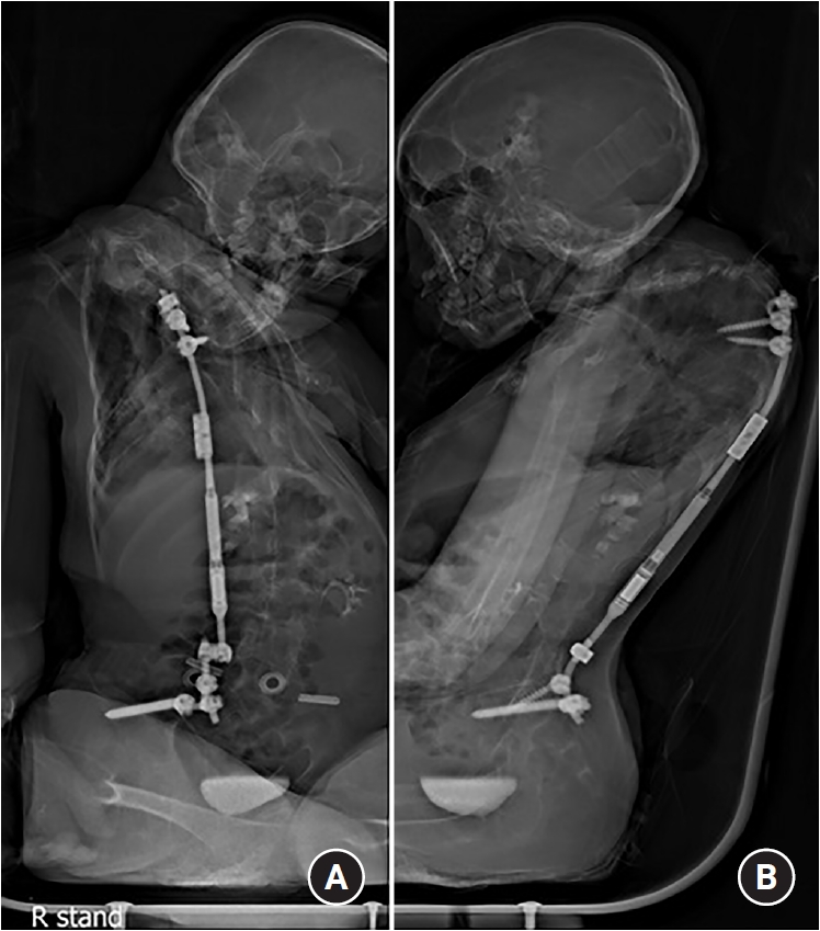
After surgery, the patient transferred to an intensive care unit for respiratory care. Mechanical ventilation was applied for one day. Frequent suctioning and chest percussion, humidified oxygen were applied four times a day. Despite these aggressive efforts, fever (38.6°C) occurred on postoperative two days. Chest X-ray showed no specific findings. However, prophylactic antibiotic (piperacillin/tazobactam) was administered. After that, no fever occurred. The patient was transferred to the general ward on postoperative three days. Whole spine X-rays performed on postoperative seven days confirmed that deformity correction had been successfully achieved. (Fig. 3) The patient's breathing was much better than before surgery.

(A) Postoperative whole spine anterior-posterior radiograph. (B) Postoperative whole spine lateral radiograph.
DISCUSSION
Morbidity following spinal deformity surgery for NSD is high, especially in respiratory complications1,4,8-10). Therefore, perioperative intensive lung care is essential for improving surgical outcome. The previous study suggests a perioperative protocol for optimizing respiratory function7). Preoperatively, it is recommended to start regimen, including percussion vest therapy, cough assist, an increase in nebulizer treatments, and frequent suctioning. Postoperatively, patients without a history of pneumonia or a tracheostomy follow a protocol that includes humidified oxygen and nebulized albuterol treatment four times per day.
NSD patients with tracheostomies were more likely to experience respiratory complications, although these preventative and aggressive regimens were applied preoperatively and postoperatively. The presence of tracheostomy is associated with negative outcomes after treatment of various conditions3,5). History of pneumonia is a clinically important independent predictor of increased risk of respiratory complications7). The association between tracheostomy presence and postoperative pneumonia has been reported3,6). The previous study reported the association of gastrostomy tube and respiratory complications7). The presence of a gastrostomy was a risk factor of respiratory complications after spinal deformity surgery in NSD patients. The presence of tracheostomy, history of pneumonia, and the presence of gastrostomy may lead to worsened surgical outcomes.
In our case, surgical correction was offered to stop the deformity progression and to maintain the function of breathing. Before surgery, bronchoscopy was done to evaluate lung condition. After the operation, the patient was transferred to intensive care unit even though there were no respiratory events during the surgery. Frequent chest percussion and suctioning, cough assist, and nebulizer treatments were applied. However, the patient developed a fever (38.6°C) on postoperative two days. Tazocin (piperacillin/tazobactam) was administered as a prophylactic antibiotic. Chest X-ray showed no specific findings, and pneumonia did not occur. Postoperative three days, the patient was transferred to a general ward. In this case, we performed aggressive lung care, including prophylactic antibiotics, before and after surgery. Given the rarity of this deformity, the optimal regimen for perioperative lung care remains unclear. However, respiratory complications can be reduced by active lung care after spinal deformity correction has been achieved in NSD patients.
CONCLUSION
A tracheostomy tube present or a history of pneumonia are risk factors of postoperative respiratory complications in NMD patients. We suggest that intensive lung care should be performed even if there were no respiratory events during the deformity correction surgery.
Notes
Conflict of interest
No potential conflict of interest relevant to this article was reported.
Acknowledgements
None.